BUILDING BLOCK 01
Beta-alanine
Beta-alanine is the less abundant partner and often helps determine how much carnosine can be synthesised in muscle. It is different from the alpha-alanine commonly used to build proteins.
Chapter one · hidden in plain sight
Its name is carnosine. It is a naturally occurring molecule, synthesised within the body and found in its highest concentrations in skeletal muscle—whether you have heard of it or not.
The molecule is tiny: just two amino acids joined together. Yet its location and unusual chemistry have kept scientists asking questions for more than a century.
Meet the molecule ↓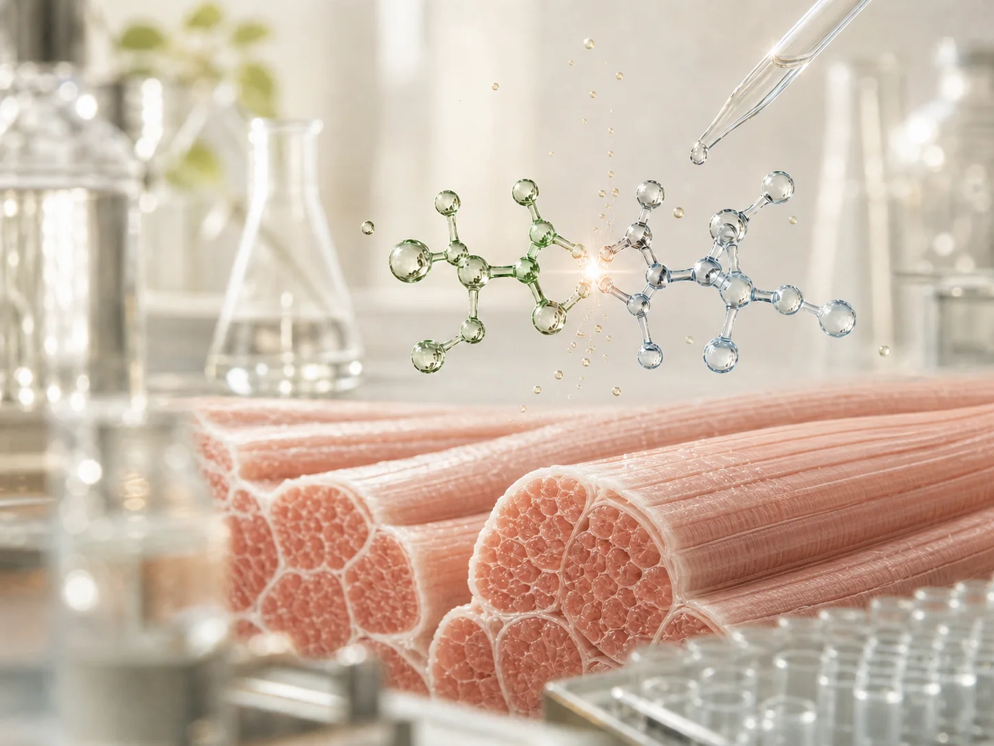
Two building blocks
one molecule found throughout muscle
First question
“Di” means two. A peptide is a short chain of amino acids. Put those ideas together and a dipeptide is simply two amino acids joined by a chemical bond.
Carnosine’s full chemical description is beta-alanyl-L-histidine. The name sounds technical; the underlying idea is not. Beta-alanine joins histidine. That single bond is the small event at the heart of this story.
So what are the two parts—and why does joining them matter? ↓
Meet the pair
Neither building block is carnosine on its own. Join them, and a distinct molecule with properties of its own comes into existence.
BUILDING BLOCK 01
Beta-alanine is the less abundant partner and often helps determine how much carnosine can be synthesised in muscle. It is different from the alpha-alanine commonly used to build proteins.
BUILDING BLOCK 02
Histidine is an amino acid used in proteins throughout the body. Its chemical structure also helps explain why researchers study carnosine in relation to acid–base chemistry.
Foundational reading: Physiology and pathophysiology of carnosine ↗
One bond creates a new molecule. The next discovery is where the body places it. ↓
Highest concentrations
skeletal muscle
Where it is found
Carnosine is synthesised when an enzyme called carnosine synthase joins beta-alanine and histidine.
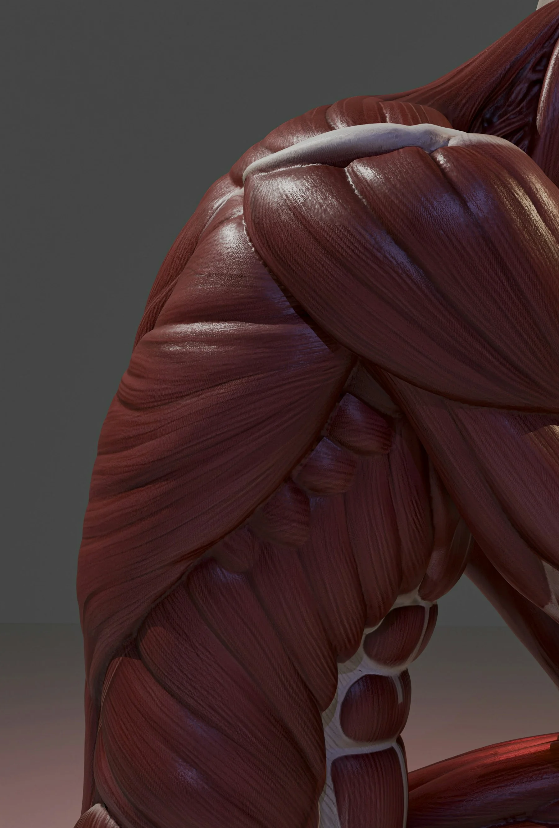
Carnosine is found in several tissues, including the brain and heart, but its concentration is highest in skeletal muscle—the muscles attached to the skeleton that help create movement.
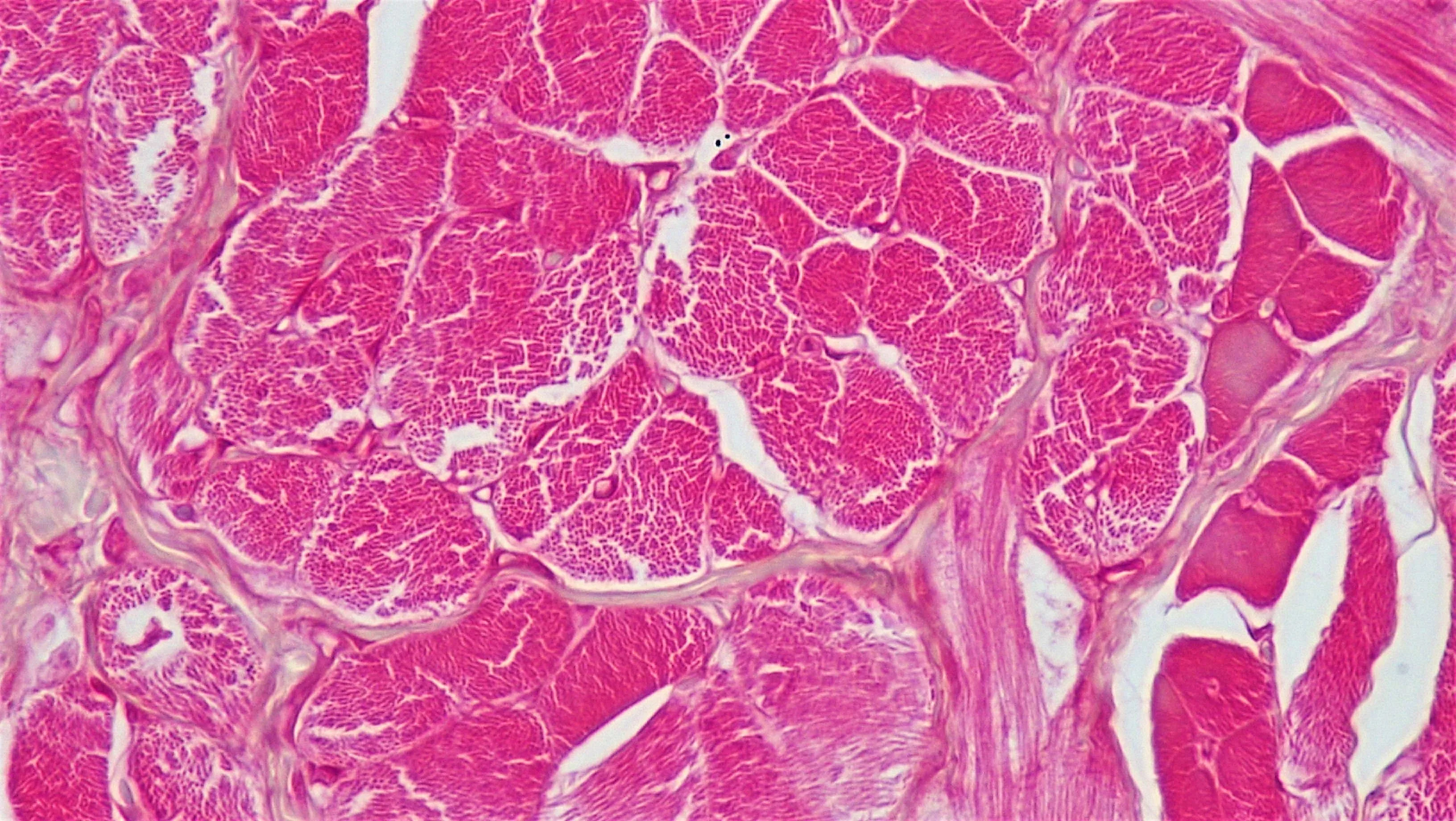
Muscle is chemically active. Its internal conditions can change quickly when it contracts. Carnosine’s abundance there, together with its chemistry, is one reason researchers have long investigated its possible physiological roles.
Why muscle?
Working muscle must continually manage chemical change. Carnosine’s structure allows it to participate in acid–base buffering—the process of moderating shifts in acidity inside cells.
This is a well-established part of why muscle researchers study carnosine. It does not mean that every proposed use of carnosine has been demonstrated, or that understanding one biological role predicts a personal outcome.
Finding a molecule in an important place tells scientists where to ask questions. It does not answer every question by itself.
Muscle explains the first century of interest. But location is only part of the story. ↓
The scientific interest
Biology provides a starting point. Research tests which explanations hold up, in which tissue and under which conditions.
Researchers study buffering and other chemical interactions in controlled systems.
Synthesis, transport and breakdown all influence how much carnosine is present in a tissue.
Tissue, measurement method and study population can all change the question being answered.
Understanding biology is not the same as making a health claim.
A chemical property, a laboratory observation and a demonstrated outcome in people are different levels of evidence.
The next question
For more than a century, researchers have explored carnosine inside the body.
So why has applying it through the skin become a scientific question?
That question begins the next chapter.
Continue the story →